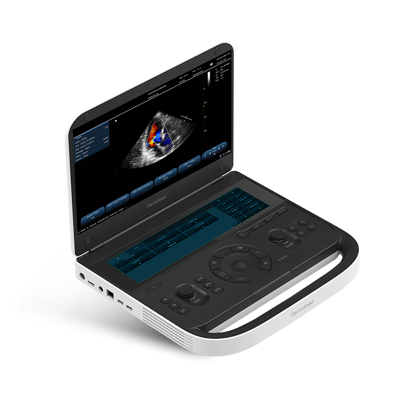
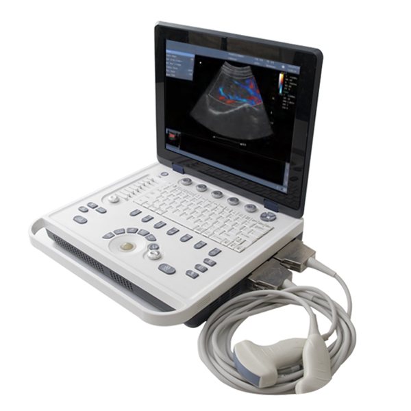
Design highlights:
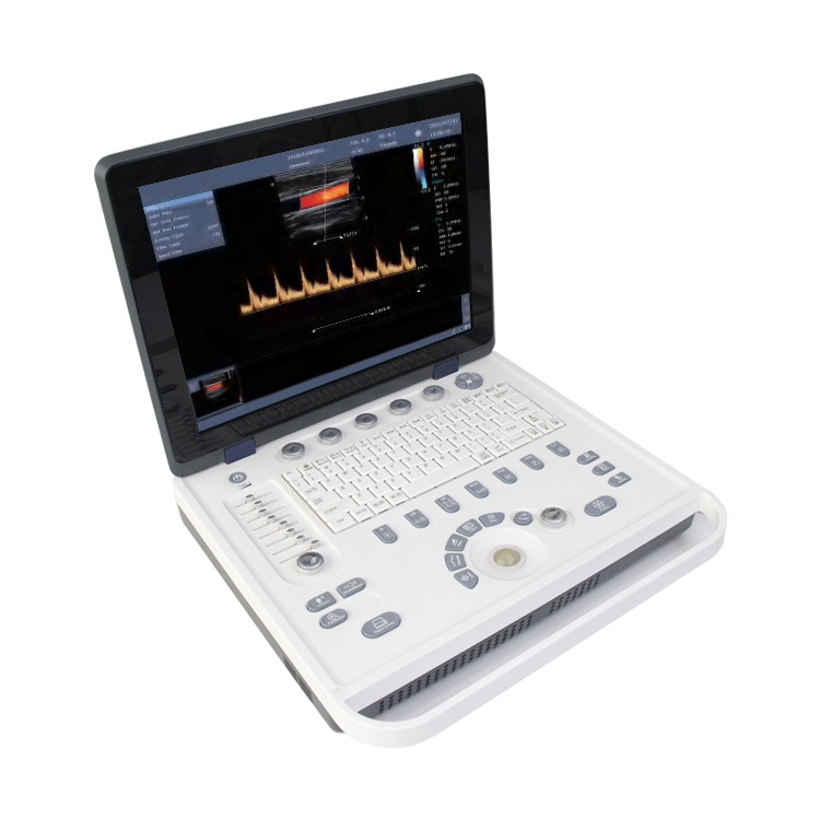
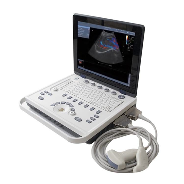
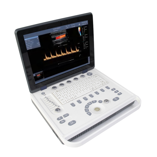
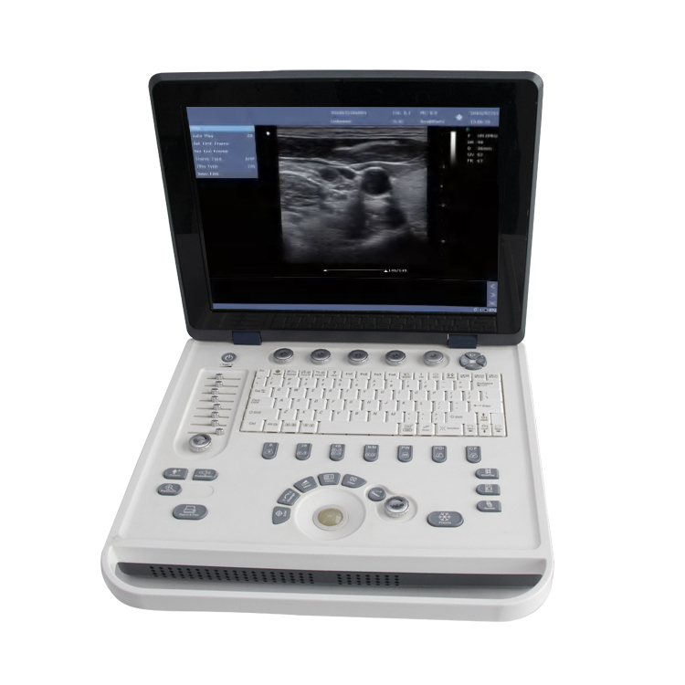
1. 15 inch medical LCD, 32 channels;
2. Built-in 500 GB hard disk for data storage;
3. Graphics and text management system to enter and classification search medical records;
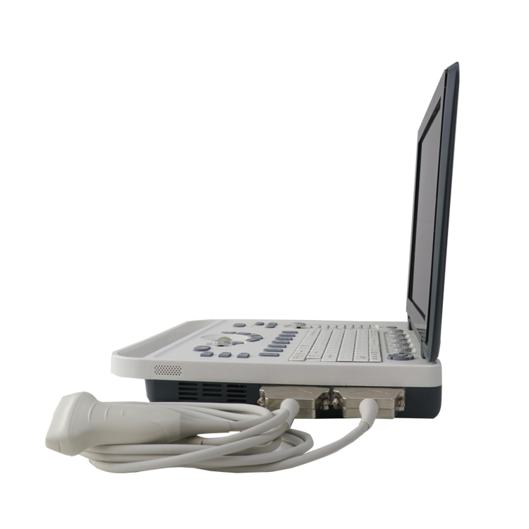
4. Notebook type with double probe interface, can be used with two probes at the same time;
5. Built-in 18650 lithium battery pack, meet the needs of daily power off use;
6. Special measurement data package for different organs;
7. Images and pathology reports can be exported.
System Imaging Function:
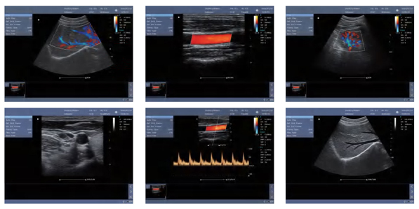
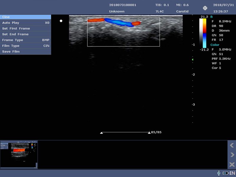
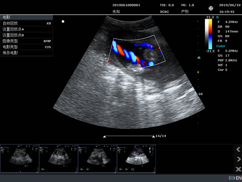
1)Color Doppler Enhancement Technology;
2)Two-dimensional grayscale imaging;
3)Power Doppler imaging;
4)PHI pulse inverse phase tissue harmonic imaging + frequency composite technique;
5)With the working mode of spatial composite imaging;
6)Linear array probe independent deflection imaging technique;
7)Linear trapezoidal spread imaging;
8)B/Color/PW trisynchronous technology;
9)Multibeam parallel processing;
10)Speckle noise suppression technology;
11)Convex expansion imaging;
12)B-mode image enhancement technique;
13)Logiqview.
Measurement and analysis:
1)General measurement: Including distance, area, circumference, volume, area ratio, distance ratio, Angle, S/D velocity, time, heart rate, acceleration, etc;
2)Obstetrics: Obstetrics supports the measurement of fetal data ≥3 fetals, including fetal weight algorithm, growth curve display, fetal echocardiography measurement (including left ventricular function measurement, left ventricular myocardial weight, etc.);
3)Fetal measurement OB1, OB2, OB3);
4)Blood flow measurement, sampling volume at least 8 levels adjustable;
5)Automatic measurement of endovascular media;
6)All measurement data Windows are removable;
7)Customized comments: Include insert, edit, save, etc.
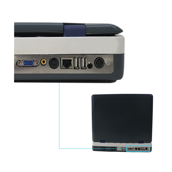
Input / output signal:
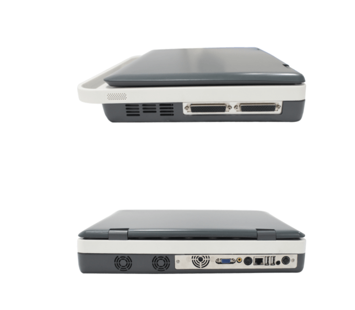
Input: Mquipped with digital signal interface;
Output: VGA, s-video,USB, audio interface, network interface;
Connectivity: Medical digital imaging and communications DICOM3.0 interface components;
Support network real-time transmission: can real-time transmission of user data to the server;
Image management and recording device: 500G hard disk Ultrasonic image archiving and medical record management function: complete;
The storage management and playback storage of patient static image and dynamic image in the host computer.
Rich data interface for data analysis:
1)VGA interface;
2)Printing interface;
3)Network interface;
4)SVIDEO interface;
5)Foot switch interface.
General system function:
1. Technology platform: linux +ARM+FPGA;
2. Color monitor: 15 " high resolution color LCD monitor;
3. Probe interface: zero force metal body connector, effectively activated two mutual common interfaces;
4. Dual power supply system, built-in large capacity lithium battery, battery power 2 hours duration, and the screen provides power display information;
5. Support quick switch function, cold start 39 seconds;
6. Main interface miniature;
7. Built-in patient data management station;8. Customized comments: include insert, edit, save, etc.
Probe Specifications:
1. 2.0-10MHz V¬ariable frequency, frequency range 2.0-10MHz;
2. 5 kinds of frequencies of each probe, variable fundamental and harmonic frequency;
3. Abdomen: 2.5-6.0MHz;
4. Superficial:5.0-10MHz;
5. Puncture Guidance: probe puncture guide is optional, puncture line and Angle are adjustable;
6. Transvaginal: 5.0-9MHZ.
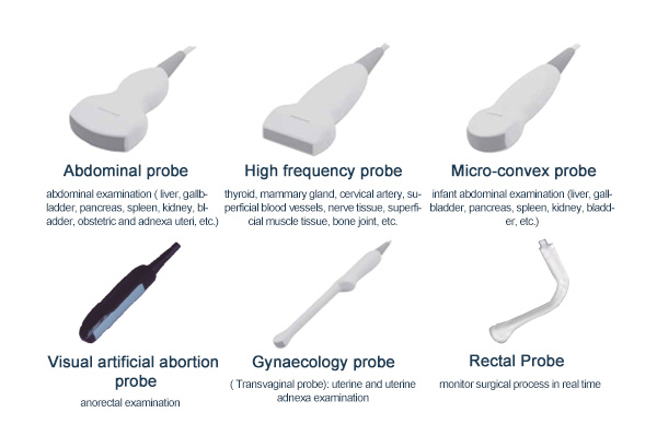
Optional Probes:
1. Abdominal probe: abdominal examination ( liver, gallbladder, pancreas, spleen, kidney, bladder, obstetric and adnexa uteri, etc.);
2. High frequency probe: thyroid, mammary gland, cervical artery, superficial blood vessels, nerve tissue, superficial muscle tissue, bone joint, etc.;
3.Micro-convex probe: infant abdominal examination (liver, gallbladder, pancreas, spleen, kidney, bladder, etc.);
4. Gynaecology probe (Transvaginal probe): uterine and uterine adnexa examination;
5. Visual artificial abortion probe: monitor surgical process in real time;
6. Rectal Probe: anorectal examination.
linux + ARM + FPGA
Probe array elements:≥ 96
3C6A: 3.5MHz / R60 / 96 array element convex probe;
7L4A: 7.5MHz / L38mm / 96 array array probe;
6C15A: 6.5MHz / R15 / 96 array element microconvex probe;
6E1A: 6.5MHz / R10 /96 array element Transvaginal probe;
Probe Frequency: 2.5-10MHz
Probe socket: 2
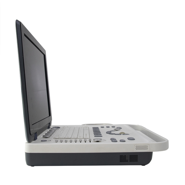
High-resolution 15-inch LCD display
Built-in 6000 mah lithium battery, working state, continuous working time for more than 1 hour, the screen provides power display information;
Supports hard drives (128GB);
Peripheral interface includes: network port, USB port (2), VGA / VIDEO / S-VIDEO, foot switch interface, support:
1.External display;
2.Video acquisition card;
3.Video printer: including black and white video printer, color video printer;
4.USB report printer: including black and white laser printer, color laser printer, color inkjet printer;
5.U disk, USB interface optical disc recorder, USB mouse;
6.foot pedal;
Host size: 370mm (length) 350mm (width) 60mm (thick)
Package size: 440mm (length) 440mm (width) 220mm (height)
Host weight: 6 kg, without probe and adapter;
Packaging weight: 10kg, (including main engine, adapter, two probes, packaging).
1.B / C mode routine measurement: distance, area, perimeter, volume, angle, area ratio, distance ratio;
2.Routine measurement of M mode: time, slope, heart rate, and distance;
3.Routine measurement of Doppler mode: heart rate, flow rate, flow rate ratio, resistance index, beat index, manual / automatic envelope, acceleration, time, heart rate;
4.Obstetrics B, PW mode application measurement: including a comprehensive obstetric radial line measurement, body weight, singleton gestational age and growth curve, amniotic fluid index, fetal physiological score measurement, etc;
5.Gynecologic B mode for applied measurement;
6.Cardiac B, M, and PW mode were applied for measurement;
7.Vascular B, PW mode application measurement, support: IMT automatic intima measurement;
8.Small organ B mode was applied measurement;
9.Urology B mode applied measurement;
10.Paediatric B mode application measurement;
11.Abdominal B mode application measurement.
standard accessories:
1.One main unit ( built-in 128G hard disk);
2.One 3C6A convex array probe;
3.Operator’s manual;
4.One power cable;
Optional accessories:
1. 6E1A Transvaginal probe;
2. 7L4A linear probe;
3. 6C15A microconvex probe;
4. The USB report printer;
5. Black and white video printer;
6. Color video printer;
7. The puncture frame;
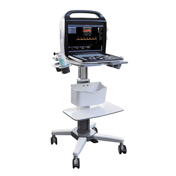
8. Trolley;
9. Foot pedal;
10.U disk and USB extension line.
1. Multi-wave beam synthesis;
2. Real-time, point-by-point, dynamic focus imaging;
3. ★ pulse reverse phase harmonic composite imaging;
4. ★ space composite;
5. ★ image-enhanced noise reduction.
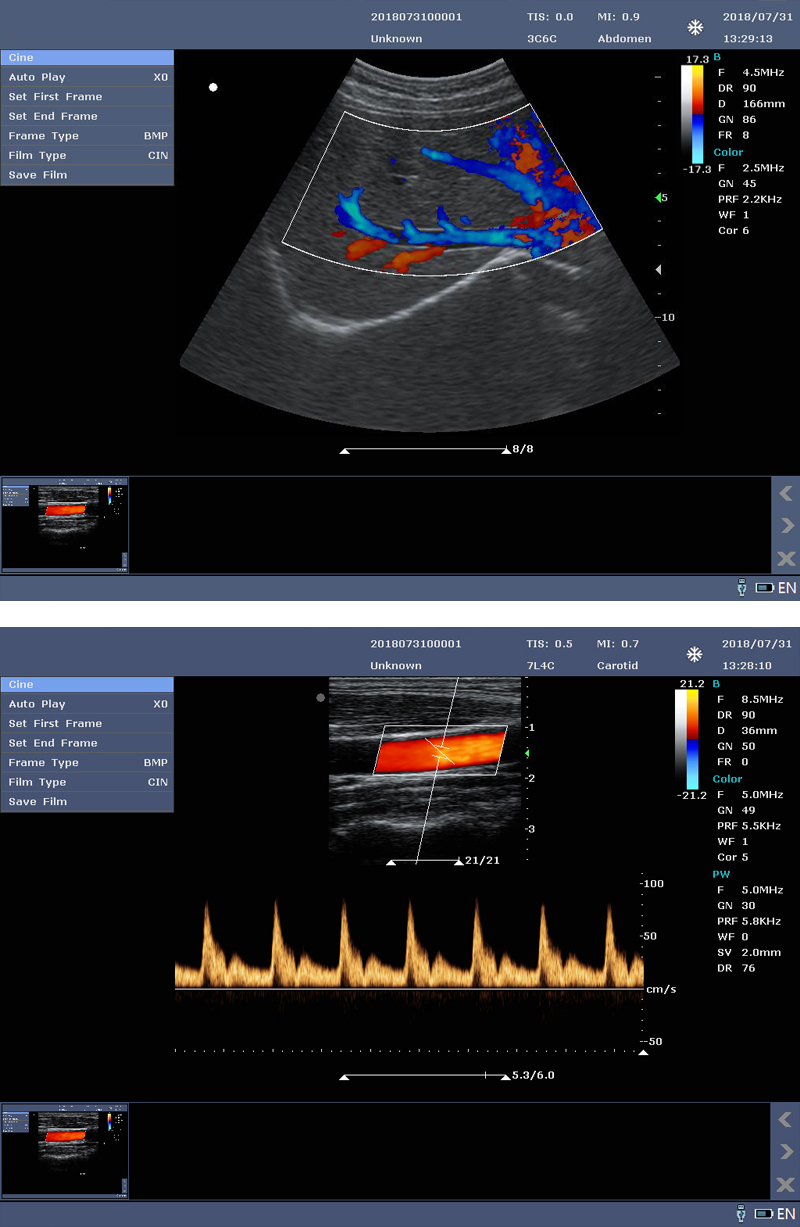
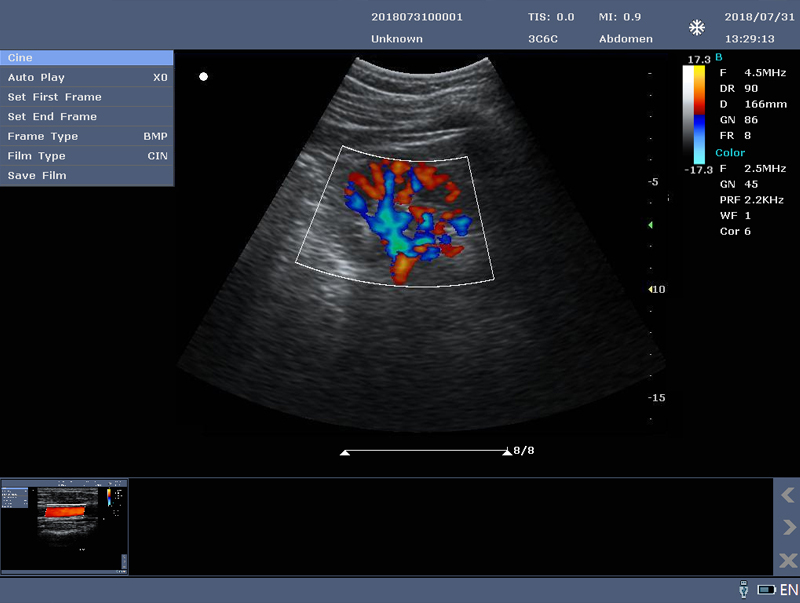
1. B mode;
2. M mode;
3. Color (color spectral) mode;
4. PDI (Energy Doppler) mode;
5. PW (pulsed Doppler) mode.
B, double, 4-amplitude, B + M, M, B + Color, B + PDI, B + PW, PW, B + Color + PW, B + PDI + PW, ★ B / BC dual real-time.
B / M: base wave frequency ≥3; harmonic frequency≥ 2;
Color / PDI ≥ 2;
PW ≥2.
1. 2D mode, B maximum≥5000 frames, Color, PDI maximum≥2500 frames;
2. Timeline mode (M, PW), maximum: 190s.
Real-time scan (B, B + C, 2B, 4B), status: infinite amplification.
1. Support for JPG, BMP, FRM image formats and CIN, AVI movie formats;
2. Support for local storage;
3. Support for DICOM, to meet the DICOM3.0 standard;
4. built-in workstation: to support patient data retrieval and browsing;
Chinese / English / Spanish / French / German / Czech, extended support for other languages according to user needs;
Abdominal, gynecology, obstetrics, urinary department, cardiac, pediatrics, small organs, blood vessels, etc.;
Support report editing, report printing, and ★ supports report template;
Annotation, landmarks, puncture line, ★ PICC, and ★ gravel line;
1. Gray scale mapping≥15;
2. Noise suppression≥8;
3. Frame correlation≥8;
4. Edge enhancement≥ 8;
5. Image enhancement≥ 5;
6. Space composite: Switch-adjustable;
7. Scan density: high, medium, and low;
8. Image flip: up and down, left and right;
9. Maximum scan depth≥320mm.
1. Scan speed (Sweep Sleep)≥5 ( adjustable);
2. Line Average (Line Average)≥ 8.
1. SV size / location: The SV size 1.0–8.0mm is adjustable;
2. PRF: 16 gear, 0.7kHz-9.3KHz adjustable;
3. Scan speed (Sweep Sleep): 5 gear is adjustable;
4. Correction Angle (Correction Angle): -85°~85°, step length of 5°;
5. Map flip: the switch is adjustable;
6. Wall filter≥ 4 gear(adjustable);
7. Polytrum sound≥20 gear.
1. PRF≥ 15 gear, 0.6KHz 11.7KHz;
2. Color Atlas (color map)≥ 4 species;
3. Color correlation≥ 8 gear;
4. Post-processing≥ 4th gear.
Support image parameters for one-key saving;
Support the one-key reset of the image parameters.
1.Quality Assureance
Strict quality control standards of ISO9001 to ensure the highest quality;
Respond to quality issues within 24 hours, and enjoy 7 days to return.
2.Warranty
All products have a 1 year warranty from our store.
3.Deliver time
Most Goods will be shipped within 72 hours after payment.
4.Three packagings to choose
You have special 3 gift box packaging options for each product.
5.Design Ability
Artwork/Instruction manual/product design according to customer’s requirement.
6.Customized LOGO and Packaging
1. Silk-screen printing logo(Min. order.200 pcs);
2. Laser engraved logo(Min. order.500 pcs);
3. Color box Package/polybag Package(Min. order.200 pcs).