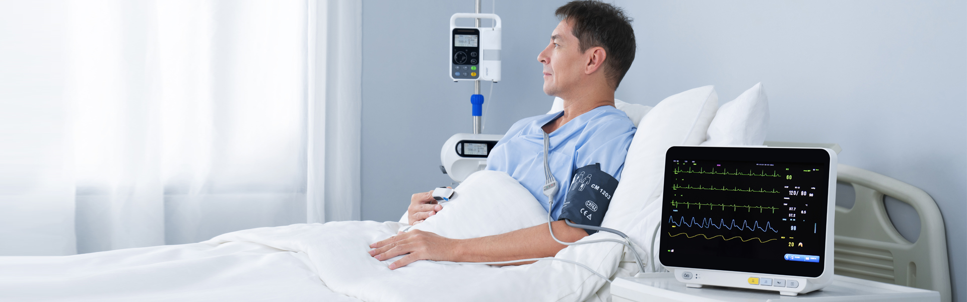
Fingertip pulse oximeter was invented by Millikan in the 1940s to monitor the concentration of oxygen in arterial blood, an important indicator of the severity of COVID-19. Yonker now explains how fingertip pulse oximeter works?
Spectral absorption characteristics of biological tissue: When light is irradiated to biological tissue, the effect of biological tissue on light can be divided into four categories, including absorption, scattering, reflection and fluorescence.If scattering is excluded, the distance that light travels through biological tissue is mainly governed by absorption. When light penetrates some transparent substances (solid, liquid or gaseous), the intensity of light decreases significantly due to the targeted absorption of some specific frequency components, which is the absorption phenomenon of light by substances. How much light a substance absorbs is called its optical density, also known as absorbance.
Schematic diagram of light absorption by matter in the whole process of light propagation, the amount of light energy absorbed by matter is proportional to three factors, which are the light intensity, the distance of the light path and the number of light-absorbing particles on the cross section of the light path. On the premise of homogeneous material, light path number light-absorbing particles on cross section can be regarded as light-absorbing particles per unit volume, namely material suction light particle concentration, can get a lambert beer's law: can be interpreted as material concentration and optical path length per unit volume of optical density, material suction light ability to respond to the nature of the material suction light.In other words, the shape of the absorption spectrum curve of the same substance is the same, and the absolute position of the absorption peak will only change due to the different concentration, but the relative position will remain unchanged. In the absorption process, the absorption of substances all takes place in the volume of the same section, and the absorbing substances are unrelated to each other, and no fluorescent compounds exist, and there is no phenomenon of changing the properties of the medium due to light radiation. Therefore, for the solution with N absorption components, the optical density is additive. The additivity of optical density provides a theoretical basis for the quantitative measurement of absorbent components in mixtures.
In biological tissue optics, the spectral region of 600 ~ 1300nm is usually called "the window of biological spectroscopy", and the light in this band has special significance for many known and unknown spectral therapy and spectral diagnosis. In the infrared region, water becomes the dominant light-absorbing substance in biological tissues, so the wavelength adopted by the system must avoid the absorption peak of water in order to better obtain the light absorption information of the target substance. Therefore, within the near-infrared spectrum range of 600-950nm, the main components of human finger tip tissue with light absorption capacity include water in blood, O2Hb(oxygenated hemoglobin), RHb(reduced hemoglobin) and peripheral skin melanin and other tissues.
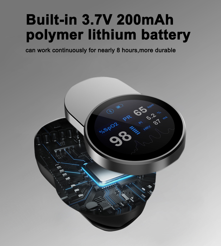
Therefore, we can obtain the effective information of the concentration of the component to be measured in the tissue by analyzing the data of the emission spectrum. So when we have the O2Hb and RHb concentrations, we know the oxygen saturation. Oxygen saturation SpO2 is the percentage of the volume of oxygen-bound oxygenated hemoglobin (HbO2) in the blood as a percentage of the total binding hemoglobin (Hb), the concentration of blood oxygen pulse so why is it called pulse oximeter? Here is a new concept: blood flow volume pulse wave. During each cardiac cycle, contraction of the heart causes blood pressure to rise in the blood vessels of the aortic root, which dilates the blood vessel wall. Conversely, diastole of the heart causes blood pressure to fall in the blood vessels of the aortic root, which causes the blood vessel wall to contract. With the continuous repetition of the cardiac cycle, the constant change of blood pressure in the blood vessels of the aortic root will be transmitted to the downstream vessels connected with it and even to the whole arterial system, thus forming the continuous expansion and contraction of the whole arterial vascular wall. That is, the periodic beating of the heart creates pulse waves in the aorta that ripple forward along the blood vessel walls throughout the arterial system. Each time the heart expands and contracts, a change in pressure in the arterial system produces a periodic pulse wave. This is what we call the pulse wave. Pulse wave can reflect many physiological information such as heart, blood pressure and blood flow, which can provide important information for non-invasive detection of specific physical parameters of human body.

In medicine, pulse wave is usually divided into pressure pulse wave and volume pulse wave two types. Pressure pulse wave mainly represents blood pressure transmission, while volume pulse wave represents periodic changes in blood flow. Compared with pressure pulse wave, volumetric pulse wave contains more important cardiovascular information such as human blood vessels and blood flow. The noninvasive detection of typical blood flow volume pulse wave can be achieved by photoelectric volumetric pulse wave tracing. A specific wave of light is used to illuminate the measurement part of the body, and the beam reaches the photoelectric sensor after reflection or transmission. The received beam will carry the effective characteristic information of the volumetric pulse wave. Because the blood volume changes periodically with the expansion and contraction of the heart, when the heart diastole, the blood volume is the smallest, blood absorption of light, the sensor detected the maximum light intensity; When the heart contracts, the volume is maximum and the light intensity detected by the sensor is minimum. In the non-invasive detection of fingertips with blood flow volume pulse wave as the direct measurement data, the selection of spectral measurement site should follow the following principles
1. The veins of blood vessels should be more abundant, and the proportion of effective information such as hemoglobin and ICG in the total material information in the spectrum should be improved
2. It has obvious characteristics of blood flow volume change to effectively collect volume pulse wave signal
3. In order to obtain the human spectrum with good repeatability and stability, the tissue characteristics are less affected by individual differences.
4. It is easy to carry out spectral detection, and easy to be accepted by the subject, so as to avoid the interference factors such as fast heart rate and measurement position movement caused by the stress emotion.
Schematic diagram of blood vessel distribution in human palm The position of the arm can hardly detect the pulse wave, so it is not suitable for the detection of blood flow volume pulse wave; The wrist is near the radial artery, the pressure pulse wave signal is strong, the skin is easy to produce mechanical vibration, may lead to the detection signal in addition to the volume pulse wave also carry skin reflection pulse information, it is difficult to accurately characterize the characteristics of blood volume change, is not suitable for measurement position; Although the palm is one of the common clinical blood drawing sites, its bone is thicker than finger, and the pulse wave amplitude of palm volume collected by diffuse reflection is lower. Figure 2-5 shows the distribution of blood vessels in the palm. Observing the figure, it can be seen that there are abundant capillary networks in the front part of the finger, which can effectively reflect the hemoglobin content in the human body. Moreover, this position has obvious characteristics of blood flow volume change, and is the ideal measurement position of volume pulse wave. The muscle and bone tissues of fingers are relatively thin, so the influence of background interference information is relatively small. In addition, the finger tip is easy to measure, and the subject has no psychological burden, which is conducive to obtain stable high signal-to-noise ratio spectral signal. Human finger consists of bone, nail, skin, tissue, venous blood and arterial blood. In the process of interaction with light, the blood volume in finger peripheral artery changes with heart beating, resulting in the change of optical path measurement. While the other components are constant in the whole process of light.
When a particular wavelength of light is applied to the epidermis of the fingertip, the finger can be regarded as a mixture, including two parts: static matter (the optical path is constant) and dynamic matter (the optical path changes with the volume of the material). When the light is absorbed by the fingertip tissue, the transmitted light is received by a photodetector. The intensity of transmitted light collected by the sensor is obviously attenuated due to the absorbability of various tissue components of human fingers. According to this characteristic, the equivalent model of finger light absorption is established.
Suitable person:
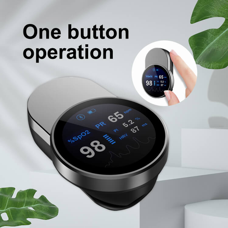
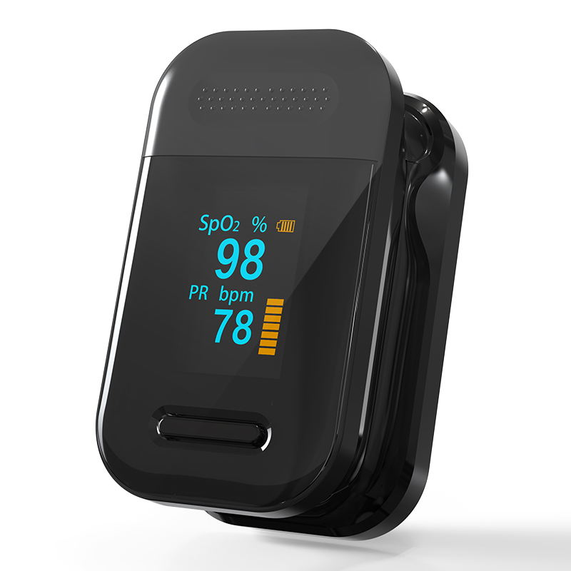
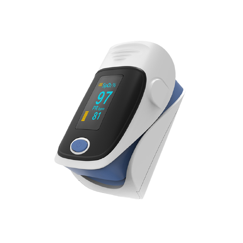
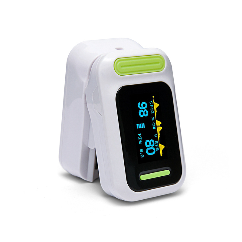
Fingertip pulse oximeter is suitable for people of all ages, including children, adults, the elderly, patients with coronary heart disease, hypertension, hyperlipidemia, cerebral thrombosis and other vascular diseases and patients with asthma, bronchitis, chronic bronchitis, pulmonary heart disease and other respiratory diseases.
Post time: Jun-17-2022